Chemotherapy remains one of the most effective treatments for breast cancer, but it often comes at a high cost for patients. Fatigue, nausea, hair loss, nerve damage, infections, and long-term cognitive effects are well-documented consequences of standard chemotherapy regimens. For patients with ERBB2-positive breast cancer, previously known as HER2-positive disease, chemotherapy is still considered essential to achieve the best clinical outcomes, even when targeted therapies are available.
A new clinical study published in JAMA Oncology suggests that an innovative immunotherapy approach delivered directly into breast tumours may help prepare the immune system to fight cancer earlier, potentially reducing reliance on intensive chemotherapy in the future. The research was led by Dr Hyo S. Han at the H. Lee Moffitt Cancer Center and Research Institute in Tampa, Florida, and represents a growing effort to harness the tumour microenvironment itself as a therapeutic target.
The study, titled “Alteration of the tumor microenvironment with intratumoral dendritic cells before chemotherapy in ERBB2 breast cancer”, explores whether immune cells injected directly into breast tumours can safely trigger anti-tumour immune responses before standard chemotherapy begins. While the trial is small and early phase, the findings offer insight into how immunotherapy may reshape breast cancer treatment strategies.
Why ERBB2-positive breast cancer still needs better options
ERBB2-positive breast cancer accounts for approximately 15 to 20 percent of all breast cancer cases and is characterized by overexpression of the ERBB2 receptor. Over the past two decades, therapies targeting this receptor, such as trastuzumab and pertuzumab, have dramatically improved survival rates. These drugs work by blocking ERBB2 signaling and by recruiting immune mechanisms to attack tumour cells.
Despite these advances, chemotherapy remains a core component of treatment. Clinical trials consistently show that combining chemotherapy with ERBB2 targeted therapy produces higher rates of pathological complete response, meaning no detectable cancer remains at the time of surgery. Achieving a pathological complete response is strongly associated with improved long-term disease-free survival.
However, chemotherapy-related toxicity continues to affect quality of life and may lead to treatment interruptions or long-term complications. This has driven interest in treatment de-escalation strategies, particularly approaches that could enhance immune-mediated tumour destruction before chemotherapy is introduced. The immune system plays a critical role in determining response to therapy, and tumours rich in immune cell infiltration are more likely to respond well to treatment.
The tumour microenvironment as a therapeutic target
Breast tumours do not exist in isolation. They are surrounded by a complex tumour microenvironment composed of immune cells, blood vessels, connective tissue and signaling molecules. In ERBB2-positive breast cancer, this microenvironment is often poorly infiltrated by immune cells and dominated by immunosuppressive factors that allow cancer to evade immune detection.
Previous research has shown that tumors with higher levels of tumor-infiltrating lymphocytes, particularly CD8-positive T cells and CD4+ T helper type 1 cells, are more likely to respond to neoadjuvant therapy. These immune cells can directly kill cancer cells or support broader immune responses that limit tumour growth.
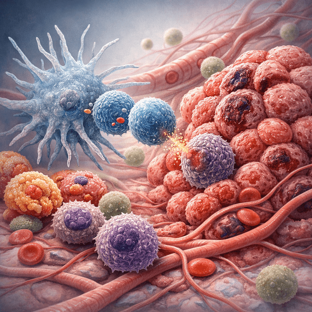
Dendritic cells play a central role in this process. They act as antigen-presenting cells, capturing tumour antigens and instructing T cells on how to recognize and attack cancer. Conventional type 1 dendritic cells, known as cDC1, are especially effective at activating cytotoxic T cell responses. In many cancers, however, these cells are scarce or functionally impaired within the tumour microenvironment.
Injecting immune teachers directly into tumours
A study conducted at Moffitt Cancer Center investigated whether autologous cDC1 cells could be generated from patients and injected directly into breast tumors prior to chemotherapy. This intratumoral delivery approach aims to bypass systemic immune suppression and stimulate immune activity precisely where the cancer resides.
Twelve patients with stage I to III ERBB2-positive breast cancer were enrolled in the phase 1 trial. All patients received intratumoral cDC1 injections once a week for six weeks, in addition to standard ERBB2-targeted therapy with trastuzumab and pertuzumab. Two dose levels of cDC1 were evaluated: 50 million cells and 100 million cells. After this immunotherapy phase, patients went on to receive paclitaxel chemotherapy before surgery.
The primary goals of the study were to assess safety and immune responses. Secondary outcomes included tumour shrinkage measured by breast magnetic resonance imaging and pathological response at surgery. Although the trial was nonrandomized and involved a small cohort, it was designed to establish whether this approach could be safely integrated into existing treatment protocols.
Safety and tolerability of intratumoral dendritic cell therapy
One of the most important findings of the study was that intratumoral cDC1 therapy was well tolerated. No dose-limiting toxicities were observed at either dose level. Most side effects were mild to moderate and included chills, fatigue, headache, and injection site reactions. These symptoms are consistent with immune activation and are commonly seen in other forms of immunotherapy.
Importantly, no severe autoimmune reactions or serious systemic toxicities were reported. This safety profile is particularly relevant when considering treatment strategies for early-stage breast cancer, where long-term toxicity must be carefully weighed against potential benefit. The higher dose of 100 million cDC1 cells was selected for further evaluation in an ongoing phase 2 trial based on its favorable safety and immunogenicity profile.
Evidence of immune activation inside the tumour
Beyond safety, the study provided compelling evidence that intratumoral cDC1 therapy altered the tumour microenvironment. Tissue biopsies taken before and after immunotherapy showed increased infiltration of immune cells within the tumour. These included CD3-positive T cells, CD8 cytotoxic T cells, CD4 helper T cells, and B cells, particularly in patients who received the higher dose of cDC1.
Advanced multiplex immunofluorescence imaging revealed that tumours initially lacking immune cells became populated with both innate and adaptive immune effectors after treatment. This shift suggests that dendritic cells injected into the tumour could recruit and activate immune cells locally.
Interestingly, while immune responses increased within the tumour, ERBB2-specific T cell responses in the blood appeared to decrease in some patients. This pattern may reflect migration of activated immune cells from the bloodstream into the tumour site and nearby lymph nodes, rather than loss of immune function.
Tumour shrinkage before chemotherapy begins
Radiological assessment using breast magnetic resonance imaging showed that most patients experienced a reduction in tumour size following the immunotherapy phase, even before chemotherapy was initiated. Eight out of twelve patients demonstrated more than a 50 percent reduction in tumour volume. Some patients showed complete or partial radiological responses at this early stage.
Circulating tumour DNA analysis provided additional evidence of tumour burden reduction. Among patients who received the higher cDC1 dose and had detectable circulating DNA at baseline, levels became undetectable after six weeks of immunotherapy. These findings suggest that intratumoral immune activation may not only affect the primary tumour but also reduce microscopic disease elsewhere in the body.
At surgery, seven of the twelve patients achieved a pathological complete response. All patients with hormone receptor-negative ERBB2-positive disease achieved a complete response, while nearly half of the hormone receptor-positive patients did so. Although chemotherapy was still administered, the early immune-mediated effects observed raise questions about how much of the overall response was driven by immunotherapy.
Reference
Han, H. S., Aldrich, A. L., Garg, S. K., Weinfurtner, R. J., Nguyen, J. V., Mo, Q., Whiting, J., Childress, J., Soliman, H., Costa, R., Armaghani, A., Soyano, A., Kiluk, J., Hoover, S., Lee, M. C., Khakpour, N., Shenoi, N., Jameel, Z., Koski, G. K., & Czerniecki, B. J. (2025). Alteration of the tumor microenvironment with intratumoral dendritic cells before chemotherapy in ERBB2 breast cancer. JAMA Oncology, 11(2), 119–127. https://doi.org/10.1001/jamaoncol.2024.5371